
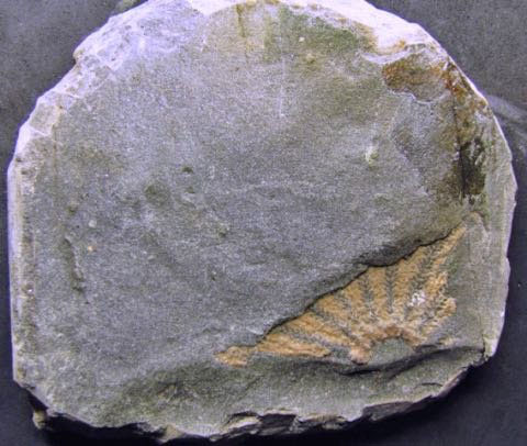
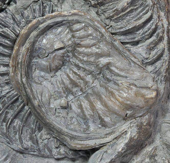
I showed you this 12.5 cm specimen of an Angulaticeras in the previous post,
but cunningly only from one side, stating it has these peculiar holes in what is left of the shell.
This specimen is wholly septate, the body chamber is completely missing, as is a part of the outer whorl.
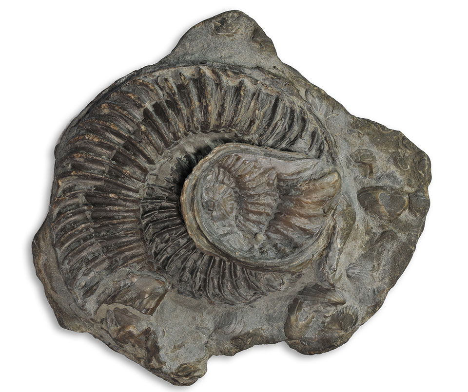
Here now both sides of the specimen, with the location of the holes marked in different
colours for the different sides.

Both sides of ammonite shown with bite marks shown in red and blue, also possible sratch marks and tear-outs
Interestingly, there seem to be roughly corresponding holes on both sides of the shell,
although this is hard to see side by side.

Picture overlay of both sides of the ammonite, showing the bite marks. Green dots showing a potential jaw alignment
Tentative alignment of the holes has been added in green dots in form of a triangular jaw
geometry (and this is of course not the only possible interpretation, as we shall see),
which would indicate the application of at least 3 bites of a marine predator
e.g. Ichthyosaur, plesiosaur, crocodile, large fish !
If one follows this interpretation, the first two possible bites probably were softer bites
to grab the ammonite, stop it from escaping while the third bite targeted the juicy
content of the body chamber, biting it off thus separating meat from shell.
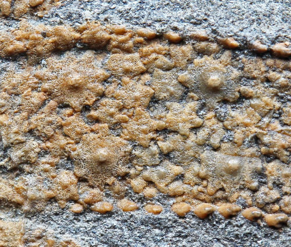
Diameters of the single holes on each of the sides vary between 4 and 8 mm,
there appear to be several combined holes from the consecutive overlapping
bites. There are no visible rims around the edges of the holes, and no apparent
shell material in the holes, but the holes seem to be fully sediment filled and could
be explored more by further preparation.

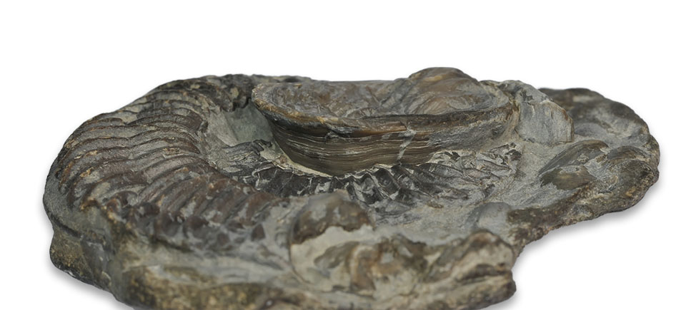
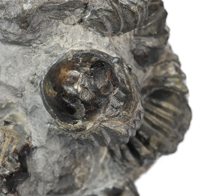
Single, double and triple bite marks

Detail of shell with double bite mark, scratch mark and bitten off shell line

Detail of bite mark with tear-out
Now there’s some very interesting literature about cretaceous ammonites showing
potential mosasaur bite marks, and some also very interesting literature showing
that these marks could also be crushed-in resting scars of patellid limpets
(literature references see below).
Kase, Seilacher et al had 1998 offered a convincing alternative explanation for the
holes to be limpet resting scars punched through by sedimentary pressure.
They had even found limpet scraping traces and limpet shells to underpin their theory,
so some significant doubt was cast over the mosasaur bite hypothesis,
as tests performed by them on nautilus shells also showed that shells were more
likely to disintegrate than be punctured when bitten.
In 2001, the mosasaur bite hypothesis was defended again (Tsujita & Westermann),
by examining a larger number of bitten shells and coming to the conclusion that,
given the rarity of limpet shells in the cretaceous Bearpaw formation of Alberta,
the mosasaur bite explanation was more likely.
In 2009, as part of an MSC thesis (D.S.King), experiments again performed on nautilus
shells, showed that punctures were very possible on the nautilus phragmocone,
that certain double holes could result from crooked teeth and that indentations
do no always necessarily have to be on both sides of a shell.
In this case however, the limpet hypothesis seems very unlikely, the holes are
relatively small, they are on the phragmocone, and there are also roughly
corresponding holes on the other side of the ammonite.
The dense lobes on this entirely septate part of the shell would probably
have provided enough structural integrity to stop this part of the ammonite
shell being broken apart in contrast to the tests having been conducted the
nautilus shells, which caused the nautilus shells to split entirely.
Other tests (King 2009) showed that holes punched into she shell are even
possible on a nautilus shell, and that it is possible that not all teeth need to
leave a mark on the shell, when the predator only administered a less powerful bite.
Interpretation as bite marks is always more interesting and headline-grabbing,
but I think in this case probability for this theory to be true is very high.
As for the question in the title of the post (who actually bit the ammonite),
I guess we can only speculate.
The bite is relatively small, the angle would probably favour a slender snout.
We know that ichthyosaurs were around, less fossil remains are known of
plesiosaurs or crocodiles in the Yorkshire lower lias.
Diameter of the holes would probably not rule out any of the predators,
so unless somebody more knowledgeable in the diagnosis of bite marks comes
up with a clever idea, the question has to remain unanswered.
The whole appearance of the Angulaticeras is extraordinarily similar to what
Kaufmann & Kesling 1960 described from cretaceous Placenticeras,
albeit this one is about 100 million years older and of course mosasaurs
did not exist then.
In my opinion this is a very likely example of reptilian bite marks on a
lower liassic ammonite and generally the oxycone shell of these types
of ammonites offers a favourable chance of preservation of these bite marks.
I had initially intended to also provide a 3D image of the ammonite,
but due to my limited spare time at the moment and the requirements
of the method (100+ pictures…) this will have to wait until a later time –
I did not want to hold posting of this blog post for much longer.
If you do have similar ammonites or want to provide feedback on this
blog post, please use the e-mail address provided in the “About me”
section of this blog.
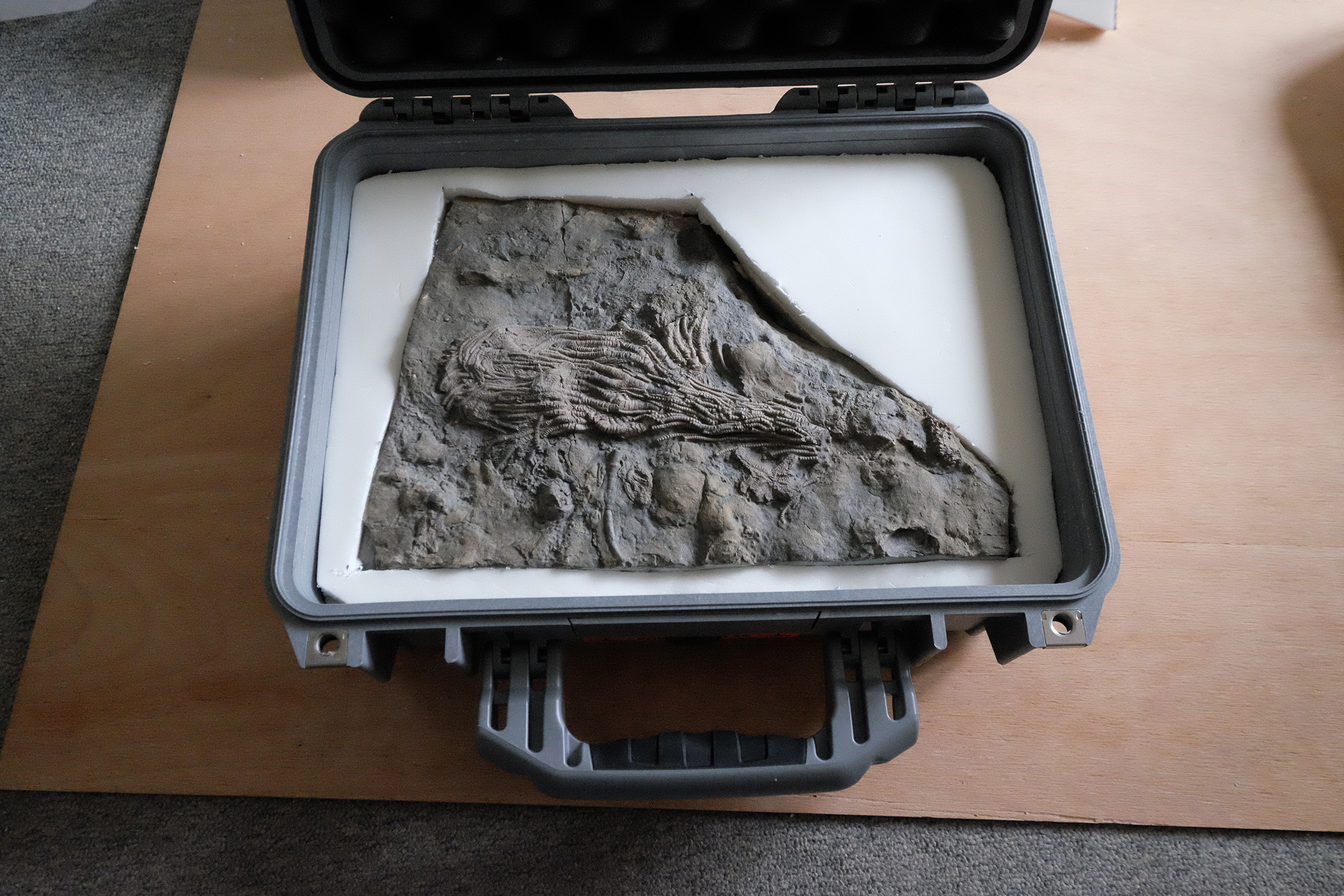
The ammonite shown is not for sale, and my intention is to donate it to a
suitable UK museum in the future.
AndyS
Literature : (links were checked at time of posting but are not guaranteed to be functional for any length of time)
-
Mosasaur bites and limpet scrapes – Wired April 11, 2012
-
Gale, A., Kennedy, W., Martill, D. 2017. Mosasauroid predation on an ammonite – Pseudaspidoceras – from the Early Turonian of south-eastern Morocco. Acta Geologica Polonica. doi: 10.1515/agp-2017-0003
-
Kauffman, E.G. and Kesling, R.V., 1960. An Upper Cretaceous ammonite bitten by a mosasaur. Contrib.Mus. Paleontol.Univ. Mich. 15:193-248
-
Kase, T., Johnston, P.A., Seilacher, A. and Boyce, J.B., 1998. Alleged mosasaur bite marks on Late Cretaceous ammonites are limpet (patellogastropod) home scars. Geology, 26(10), pp.947-950.
-
Tsujita, C.J. and Westermann, G.E., 2001. Were limpets or mosasaurs responsible for the perforations in the ammonite Placenticeras?. Palaeogeography, Palaeoclimatology, Palaeoecology, 169(3), pp.245-270.
-
Klompmaker, A.A., Waljaard, N.A. and Fraaije, R.H., 2009. Ventral bite marks in Mesozoic ammonoids. Palaeogeography, Palaeoclimatology, Palaeoecology, 280(1), pp.245-257.
-
Hewitt, R.A. and Westermann, G.E.G., 1990. Mosasaur tooth marks on the ammonite Placenticeras from the Upper Cretaceous of Alberta,Canada. Canadian Journal of Earth Sciences, 27(3), pp.469-472.
-
The Abilty of Mosasaurs to produce unique puncture marks on ammonite shells, MSC thesis Daniel Steven King, Bowling Green State University 2009


, to the right the snout of dear beach dog Lucy")


 ammonites")
